Thursday, February 28, 2013

Alagille Syndrome


Alagille Syndrome


What is Alagille syndrome?

  • Alagille syndrome is an inherited disorder that mimics other forms of prolonged liver disease seen in infants and young children. 
  •  However, a group of unusual features in other organ systems distinguishes Alagille syndrome from other liver and bile duct diseases in infants.
  • Specifically, Alagille syndrome is also associated with cardiac disease, eye and skeletal findings and a characteristic facial appearance.
  • The blood vessels and kidneys may also be involved in a smaller proportion of cases.
  • Children with Alagille syndrome usually have a liver disease characterized by a progressive loss of the bile ducts within the liver over the first year of life.
  • This leads to a buildup of bile in the liver, causing damage to liver cells. 
  • Scarring may occur and lead to cirrhosis in about 30 to 50 per cent of affected children.


What causes Alagille syndrome?

  1. Alagille syndrome is caused by changes, or mutations, in one of two genes, usually JAGGED1 or occasionally NOTCH2.
  2.  In approximately 60 per cent of the cases of Alagille syndrome, the gene change is the result of a new mutation that occurs in that baby.
  3. In other 40 per cent of cases, the gene mutation is inherited from one of the parents.
  4.  A parent carrying a disease-causing mutation has a 50 per cent chance of transmitting that gene to any subsequent children.
  5.  Each affected adult or child may have all or only a few of the features of the syndrome. 
  6. Even individuals in the same family, sharing the same gene mutation have variable manifestations in each of the affected organ systems.


What are the symptoms and physical characteristics of Alagille syndrome?

  • Not all individuals with Alagille syndrome have significant liver disease, though probably the majority do.
  • Typically, symptoms of the illness are jaundice and poor growth within the first three months of life. 
  • Later, there is persistent jaundice, severe itching, fatty deposits in the skin (xanthomas) and poor growth during early childhood. 
  • Frequently the disease stabilizes between ages five and eight with an improvement in symptoms.

  • Other features which help establish the diagnosis include abnormalities in the cardiovascular system, the spinal bones, the eye and the kidneys. 
  • Heart involvement typically involves narrowing of the blood vessel connecting the heart to the lungs (pulmonary artery) which ranges in severity, but may also include a simple heart murmur or a severe congenital heart problem.
  • The shape of the bones of the spinal column may often look like the wings of a butterfly on x-ray, but this does not cause any problems.
  • There is also a specific facial appearance shared by children with Alagille syndrome that makes them easily recognizable. 
  • The features include a prominent, broad forehead, deep-set eyes, a straight nose and a small pointed chin.

The majority of children with Alagille syndrome have an abnormality of the eyes in which an extra, circular line on the surface of the eye can be detected by a specialized eye examination. 
In addition, some children have various abnormalities in their kidneys.
 In approximately 15 per cent of individuals with Alagille syndrome there is a risk of bleeding in the head and brain (stroke).


How is Alagille syndrome diagnosed?

  • Although Alagille syndrome was first described in the English medical literature in 1975, it is now becoming recognized more frequently among children with chronic forms of liver disease. 
  • Diagnosis can be established by microscopic examination of liver biopsy specimens, a stethoscope examination of the child’s heart and chest, a special eye examination (slit-lamp exam), an x-ray of the spinal column and an ultrasound examination of the abdomen.
  • Genetic testing can also be performed to confirm unusual or mild cases.


How is Alagille syndrome treated?

  • Treatment of Alagille syndrome is primarily medical and not surgical and is based on trying to increase the flow of bile from the liver, maintain normal growth and development, and prevent or correct any of the specific nutritional deficiencies that often develop. 
  •  Because bile flow from the liver to the intestine is slow in Alagille syndrome, medications designed to increase the flow of bile are frequently prescribed. 
  • This may decrease the damage in the liver and improve the digestion of fat and fat soluble vitamins.
  • Itching caused by the buildup of bile in the blood and skin may be relieved by medications (e.g. ursodeoxycholic acid, cholestyramine). 
  • Elevations in blood cholesterol also respond to the medications used to increase bile flow. 
  • Elevated blood cholesterol levels can lead to small yellow deposits of cholesterol on the skin of knees, elbows, palms, eyelids and other surfaces that are frequently rubbed (xanthomas). 
  • Although reduced flow of bile into the intestine leads to poor digestion of dietary fat, a specific type of fat can still be well digested and therefore infant formulas containing high levels of medium-chain triglycerides (MCT) are usually substituted for conventional formulas in infants. 
  • Some infants can grow adequately on breast milk if additional MCT oil is given.  Foods containing high fat levels may lead to looser, greasy stools later in childhood. 
  • However, the benefits from the calories and vitamins in the fat that is absorbed usually leads to the recommendation that the child not be put on a low-fat diet.  There are no other dietary restrictions. 
  • Occasionally, MCT oil is also prescribed as a nutritional supplement in infancy.
  • Problems with fat digestion and absorption may lead to a deficiency of fat-soluble vitamins (A, D, E and K). 
  • Vitamin A deficiency causes night blindness and red eyes. 
  • Vitamin D deficiency causes softening and fractures of the bones and teeth.  Vitamin E deficiency causes a disabling disease of the nervous system and muscles, and vitamin K deficiency causes bleeding problems and has been associated with poor bone health as well.
  • Deficiencies of these vitamins can be diagnosed by blood tests and usually can be corrected by vitamin supplements.
  • There is presently no procedure that can correct the loss of the bile ducts within the liver.
  • In 20 to 30 per cent of patients, liver cirrhosis advances to a stage where the liver fails to perform its functions. 
  •  Liver transplantation is then considered.
  • The overall life expectancy for children with Alagille syndrome is unknown, but depends on several factors: the severity of scarring in the liver and/or the need for liver transplantation, the risk of stroke and whether heart or lung problems develop because of the narrowing in the pulmonary artery. 
  • Many adults with Alagille syndrome are leading normal lives.

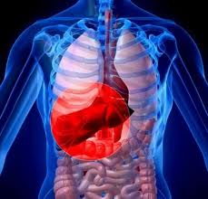
LIVER DISEASE


LIVER DISEASE
What is Liver Disease ? 


  • Liver disease is any disturbance of liver function that causes illness. 
  • The liver is responsible for many critical functions within the body and should it become diseased or injured, the loss of those functions can cause significant damage to the body. Liver disease is also referred to as hepatic disease.
  • Liver disease is a broad term that covers all the potential problems cause the liver to fail to perform its designated functions. 
  • Usually, more than 75% or three quarters of liver tissue needs to be affected before decrease in function occurs.
  •  Although liver disease is stereotypically linked to alcohol or drugs, the truth is that there are over 100 known forms of liver disease caused by a variety of factors and affecting everyone from infants to older adults. 
  • For information on specific forms of liver disease, click on 'Types of Liver Disease' in the menu on the left.


Cirrhosis 

Cirrhosis is often considered to be a form of liver disease and may be the only liver-related condition that many people have heard of. In fact, cirrhosis is a condition that results from permanent damage or scarring of the liver. It is the end stage of many different forms of liver disease and is known to cause a number of other health problems, including variceal bleeding, ascites and hepatic encephalopathy.
Many types of liver disease still have unknown causes but the most frequent liver diseases are generally caused by one of the following factors:

Viral hepatitis

  • Caused by viruses that attack the liver, viral hepatitis comes in many forms. The most common forms world-wide are hepatitis A, B and C. 
  • Although hepatitis A and B can be prevented by vaccine, there is no vaccine for hepatitis C.
  • In Canada, hepatitis C is the leading cause of liver transplants.


Obesity

The leading cause of liver disease in Canada is fatty liver disease linked to obesity.

Alcohol

Factors such as gender, age, nationality, weight and health can affect how a person’s liver metabolizes alcohol. When the liver has too much alcohol to handle, normal liver function may be interrupted leading to a chemical imbalance. If the liver is required to detoxify alcohol continuously, liver cells may be destroyed or altered resulting in fat deposits (fatty liver) and more seriously, either inflammation (alcoholic hepatitis) and/or permanent scarring (cirrhosis). Liver cancer can also result from alcohol induced liver disease.

Genetics

Several forms of liver disease are caused or thought to be caused, by defective genes. These forms of liver disease may be diagnosed in infancy or may not show up until later in life. Examples include hemochromatosis, Wilson disease,tyrosinemia, alpha 1 antitrypsin deficiency and Glycogen Storage disease.


Autoimmune disorders

Sometimes a body’s immune system may begin to attack the liver or bile ducts causing inflammation and scarring which leads to a progressive form of liver disease. Examples of liver diseases believed to be caused by the immune system are primary biliary cirrhosis (PBC), primary sclerosing cholangitis (PSC) and autoimmune hepatitis.


Drugs and toxins

The liver is responsible for processing most of the chemicals and medications that enter your body – this leaves it vulnerable to acute or chronic liver disease caused by chemicals. In some cases, this is a predictable consequence of overexposure or over-consumption of certain chemicals such as acetaminophen or industrial toxins like polyvinyl chloride or carbon tetrachloride. In other cases, chemicals can cause an unpredictable reaction.


Cancer

Although primary liver cancer is relatively uncommon, many other forms of cancer often metastasize in the liver. Because the liver filters a high volume of blood which may be carrying cancer cells, it is susceptible to developing a form of secondary cancer. If cancer originates in the liver, it is often caused by hepatitis B, hepatitis C or it can develop in cases of advanced liver disease when cirrhosis is present.

Wednesday, February 27, 2013

Brain Tumours Types


Sorting brain tumours into different types


There are nearly 100 different types of brain tumours. They are generally named after the type of cell they developed from. Most brain tumours develop from the cells that support the nerve cells of the brain called glial cells. A tumour of glial cells is a glioma.
Brain tumours can also be named after the area of the brain they are growing in. A tumour of the pituitary gland is called a pituitary adenoma. A tumour developed from the covering of the brain (the meninges) is called a meningioma. Tumours growing from the nerves entering the brain are called neuromas. An acoustic neuroma is a tumour growing on the nerve that controls hearing.

Brain tumour grade - benign or malignant

Brain tumours are put into groups according to how quickly they are likely to grow. There are 4 groups, called grades 1 to 4. The cells are examined under a microscope. The more normal they look, the more slowly the brain tumour is likely to develop and the lower the grade. The more abnormal the cells look, the more quickly the brain tumour is likely to grow and the higher the grade. Low grade gliomas (grade 1 and grade 2) are the slowest growing brain tumours.
You may have been told you have a benign tumour or a malignant tumour. As a rule of thumb, low grade tumours are regarded as benign and high grade as malignant. By benign, we generally mean the following.
·     The tumour is relatively slow growing
·     It is less likely to come back if it is completely removed
·     It is not likely to spread to other parts of the brain or spinal cord
·     It may just need surgery and not radiotherapy or chemotherapy as well
By malignant, we generally mean the following.
·     The tumour is relatively fast growing
·     It is likely to come back after surgery, even if completely removed
·     It may spread to other parts of the brain or spinal cord
·     It can't just be treated with surgery and will need radiotherapy or chemotherapy to try to stop it from coming back
With other types of cancer, these black and white explanations of benign and malignant work well. But with brain tumours, there are a lot of grey areas. Some low grade astrocytomas can spread to other parts of the brain or spinal cord. Radiotherapy and chemotherapy are sometimes used to treat benign tumours. Even a slow growing tumour can cause serious symptoms and be life threatening if it is in a crucial part of the brain. So, it is important to ask your specialist to explain your own situation to you fully and simply.

Gliomas

About half of all primary brain tumours are gliomas. There are 3 main types of glioma - astrocytoma,ependymoma and oligodendroglioma. A fourth type, mixed glioma, is a mixture of the other types. Your doctor will use the grade of your glioma to decide your treatment and the likely outcome. But the position of the tumour is also very important. For example, brain stem gliomas are particularly difficult to treat, whatever their grade. The brain stem is a very complicated and delicate part of the brain and completely removing the tumour is not likely to be possible. Unfortunately, high doses of radiotherapyare not recommended either as this may cause too much damage to the normal brain stem.

Astrocytomas (including glioblastoma multiforme)

Astrocytomas are the most common type of glioma in both adults and children. They develop from cells called astrocytes. The astrocytes are the 'bricks and mortar' of the brain that support the nerve cells (neurones). They probably do other things too (but we don't know what as yet). Astrocytomas can be slow (low grade) or fast growing (high grade). Some are very localised (focal). This means it is easy to see the border between tumour and normal brain tissue on a scan or during an operation. Focal astrocytomas are more often diagnosed in children and are not common in adults. Other astrocytomas are called diffuse. These do not have a clear boundary between the tumour and normal brain tissue.
Anaplastic astrocytoma (also called grade 3 astrocytoma) and glioblastoma multiforme (GBM or grade 4 astrocytoma) are the most common type of  brain tumour in adults. These are malignant (high grade) brain gliomas. They can sometimes spread to other parts of the brain.
Ependymomas
About 1 in 20 brain tumours (5%) is an ependymoma. These develop from cells called ependymal cells. These cells line the fluid filled areas of the brain (the ventricles) and spinal cord. Their job is to repair any damaged nerve tissue. Most ependymomas are diagnosed in children or young adults. They can be high or low grade, but the cells' appearance under a microscope does not always fit with their behaviour. So the grade may not tell you much. Sometimes ependymomas can spread within the central nervous system, via the fluid that circulates round the brain and spinal cord, but this is not common.

Oligodendrogliomas

About 1 in 20 brain tumours (5%) is an oligodendroglioma. These develop from cells called oligodendrocytes. These cells make a white fatty substance that covers nerves, called myelin. It helps the nerve signals (impulses) to travel along the nerves more quickly. Oligodendrogliomas are most often found in the forebrain, in the temporal or frontal lobes. They can be fast or slow growing. They are most likely to be diagnosed in adults, although they do occur in young children. Sometimes this tumour can spread within the central nervous system, in the fluid that circulates round the brain and spinal cord.

Mixed gliomas

These are gliomas that are a mixture of 2 or even 3 of the different types of glioma. The cell types can be different grades too. Your doctor will look at the different types of glioma cells and give you the treatment that is appropriate for the most aggressive cell type in your brain tumour.

Acoustic neuromas

Acoustic neuromas grow in the nerve that runs from the ears to the brain and controls hearing and balance. They are nearly always slow growing, do not spread and are thought of as benign brain tumours. Often, they have been there a long time by the time they are diagnosed. They are found most often in older people. Loss of hearing in one ear can be a sign of acoustic neuroma. Rarely, they are associated with one form of a genetic condition called neurofibromatosis type 2 (NF 2). In these cases, they are usually diagnosed at a much younger age, can be on both sides (bilateral) and people affected may also develop meningiomas.

Craniopharyngiomas

Craniopharyngiomas are tumours that tend to grow near the base of the brain, just above the pituitary gland. They are most often diagnosed in children, teenagers and young adults. They do not usually spread, but are near important structures in the brain and can cause problems as they grow. They can cause changes in hormone levels and problems with eyesight. Children with craniopharyngioma can have weight gain and growth problems.
 Haemangioblastomas
Only 2 out of every 100 brain tumours (2%) are haemangioblastomas. They grow from blood vessel cells. They are very slow growing and do not spread. But they can grow in the brain stem and then they are very difficult to treat. Sometimes these brain tumours can be part of a rare syndrome called von Hippel Lindau syndrome (vHL), which runs in families. People with vHL who develop haemangioblastoma may have more than one. Not everyone with vHL will get haemangioblastomas - the condition behaves differently in different people.

Lymphomas

Sometimes lymphoma can start in the brain. This is called primary cerebral lymphoma or primary central nervous system (CNS) lymphoma. Most are a type of lymphoma called diffuse large B cell non Hodgkin's lymphoma. People who have poor immunity due to AIDS or due to medicines taken after an organ transplant are more likely to develop cerebral lymphoma than other people in the population.
Lymphoma is a cancer of the lymphatic system. These tumours are treated differently to other types of brain tumour. There is a section about  non Hodgkin's lymphoma on this website, including treatment information.
Meningiomas
About 1 in 4 brain tumours in adults (25%) is a meningioma. They are more common in older people and in women. These tumours start in the tissues covering the brain (membranes). They are most often found in the forebrain or hindbrain. They are usually benign (not cancerous).
Some meningiomas are atypical. This means that they behave more aggressively than normally expected for meningiomas. They can grow into surrounding brain tissue and may come back after they have been removed.
Meningioma symptoms vary a lot, depending on where in the brain they are growing.

Germ cell tumours

A germ cell tumour grows from primitive developing cells that form in the embryo and develop into the reproductive system. Most occur outside the brain, in the chest or abdomen but they can occur in the brain. They are most commonly found in the pineal and suprasellar areas. Germ cell tumours account for about 2 out of 100 (2%) of all brain tumours in children. Around half of these tumours occur in young people between 10 and 20 years old.
Germ cell tumours sometimes produce chemicals that can be tested for in the blood - AFP and HCG. So sometimes these tumours can be diagnosed with a blood test. They are often picked up when they are still small, because they can block the circulation of fluid around the brain and tend to cause symptoms early on.

Pineal region tumours

The pineal gland is in the middle of the brain, just behind the top of the brain stem. It makes the hormone melatonin. Pineal tumours are rare and account for only 1 in every 100 brain tumours (1%). Several different types of tumours can grow in the pineal region, including gliomas. The most common types are germ cell tumours.

Pituitary tumours

About 1 in 10 brain tumours (10%) are in the pituitary gland. They are more common in older people. The pituitary gland is attached to the underside of the brain. It lies in a small hollow in the skull, just behind the eyes. It controls many body functions by making and releasing chemical messengers (hormones) into the bloodstream. The pituitary hormones travel in the blood to other glands in the body, such as the thyroid, ovaries and testicles. The pituitary hormones control amounts of other hormones that these glands release into the blood.
Most pituitary tumours develop from the gland tissue and are called adenomas. They are nearly always benign - they grow slowly and do not spread. Pituitary tumours can often cause quite odd symptoms because the tumour cells make too much of one of the pituitary hormones.

Primitive neuroectodermal tumours (PNETs)

These tumours develop from cells that are left over from the earliest development of the body in the womb. Normally, these cells are harmless. But sometimes they can become cancerous. 
Medulloblastoma is the commonest type of PNET. These grow in the hindbrain They are the second most common brain tumour in children, but the commonest malignant (high grade) childhood brain tumour. Medulloblastomas are also diagnosed in young adults.These tumours can be fast growing and can spread to other parts of the brain and to the spinal cord through the cerebrospinal fluid (CSF).

Spinal cord tumours

Up to 1 in 5 central nervous system tumours (20%) are in the spinal cord. There are a few different types. The success of treatment depends on the type of spinal cord tumour.
Meningiomas and neurofibromas are the commonest types seen in adults. They grow outside the spinal cord, but press on it. Astrocytomas and ependymomas grow in the spinal cord tissue itself. These are the commonest types in children. Another rare type is called a chordoma.
Tumours growing in the bones of the spine can press on the spinal cord and cause similar symptoms to spinal cord tumours. In adults, these are usually secondary cancers that have spread into the spinal bones from somewhere else in the body. For example, the lungs, prostate, kidney or the breast. Lymphomas and myeloma can also spread to the spine.

Monday, February 25, 2013

CONGENITAL ANOMALIES


CONGENITAL ANOMALIES


Definition

  • Congenital anomalies are also known as birth defects, congenital disorders or congenital malformations. 
  • Congenital anomalies can be defined as structural or functional anomalies, including metabolic disorders, which are present at the time of birth.

Causes and risk factors

  • Approximately 50% of all congenital anomalies, however, cannot be assigned to a specific cause. 
  • However some causes or risk factors have been associated to congenital anomalies.

Socioeconomic factors

  • Although it may be an indirect determinant, congenital anomalies are more frequent among resource constrained families and countries.
  •  It is estimated that about 94% of serious birth defects occur in middle- and low-income countries, where mothers are more susceptible to macronutrient and micronutrient malnutrition and may have increased exposure to any agent or factor that induces or increases the incidence of abnormal prenatal development, particularly infection and alcohol. 
  • Advanced maternal age also increases the risk of some chromosomal abnormalities including Down syndrome.

Genetic factors

  • Consanguinity (relationship by blood) increases the prevalence of rare genetic congenital anomalies and nearly doubles the risk for neonatal and childhood death, intellectual disability and serious birth anomalies in first cousin unions. 
  • Some ethnic communities, e.g. Ashkenazi Jews or Finns, have comparatively high prevalence of rare genetic mutations, leading to a higher risk of congenital anomalies.

Infections
  • Maternal infections such as syphilis and rubella are a significant cause of birth defects in low- and middle-income countries.

Maternal nutritional status

  • Iodine deficiency, folate insufficiency, overweight, or conditions like diabetes mellitus are linked to some congenital anomalies.
  • For example folate insufficiency increases the risk of having a baby with neural tube defects.

Environmental factors

  • Maternal exposure to pesticides, medicinal and recreational drugs, alcohol, tobacco, certain chemicals, high doses of vitamin A during the early pregnancy, and high doses of radiation increase the risk of having a baby with congenital anomalies.
  •  Working or living near or in waste sites, smelters, or mines may also be a risk factor.

Prevention

  • Preventive public health measures administered through pre- and peri-conception and prenatal health care services decrease the frequency of certain congenital anomalies. 
Primary prevention of congenital anomalies involves:

  •          Improving the diet of women throughout their reproductive years, ensuring an adequate dietary intake of vitamins and minerals such as folic acid and iodine, and restricting harmful substances, particularly the abuse of alcohol. 
  •     Controlling pre-conceptional and gestational diabetes through counselling, weight management, diet and the administration of insulin when needed.
  •          Avoiding exposure to hazardous environmental substances (e.g. heavy metals, pesticides, some medicinal drugs) during pregnancy.
  •          Improving vaccination coverage, especially with rubella virus, for children and women. 
  •     This can be prevented through childhood vaccination. The rubella vaccine can also be given at least 1 month prior to pregnancy to women who are not already immune.
  •          Increasing and strengthening education to health staff and others interested in promoting birth defects prevention.

Detection

  • Pre- and peri-conceptional care includes basic reproductive health practices as well as medical genetic screening. Screening can be conducted during the following three periods:
  •  Preconception screening is used to identify persons at risk for specific disorders or at risk for passing one on to their children. 
  • The strategy includes the use of family histories and carrier screening, and is particularly valuable in countries where consanguineous marriage is common.
  •   Antenatal screening includes screening for advanced maternal age, Rhesus blood group incompatibility, and carrier screening.
  • Ultrasound can be used to detect Down syndrome during the first trimester and serious fetal anomalies during the second trimester; maternal serum screening can also be used for detection of Down syndrome and neural tube defects during the first and second trimesters.
  • Newborn screening includes clinical examination and screening for haematological, metabolic, and hormonal disorders. 
  • Screening for deafness and heart defects as well as early detection of birth defects can facilitate life-saving treatments and prevent the progression towards some physical, intellectual, visual or auditory disabilities.

Treatment and care

  • In countries with well-established health services, structural birth defects can be corrected with paediatric surgery and early treatment can be administered to children with functional problems such as thalassaemia (inherited recessive blood disorders), sickle cell disorders and congenital hypothyroidism.

WHO response

  • In 2010, the World Health Assembly issued a report on birth defects. The report describes the basic components for creating a national programme for the prevention and care of birth defects before and after birth. It also recommends priorities for the international community to assist in establishing and strengthening of these national programmes.
  • The Global Strategy for Women’s and Children’s Health, launched in 2010 by the UN in collaboration with leaders from governments and other organizations like WHO and UNICEF, has been crucial in implementing high-impact and cost-effective interventions to improve neonatal and child health.
  • WHO is also working with the US Centers for Disease Control and Prevention’s (CDC) National Center on Birth Defects and Developmental Disabilities and other partners to establish a global policy for folate fortification at the country-level and to provide needed technical expertise for the surveillance of neural tube defects, for monitoring folic acid fortification efforts and improve laboratory capacity on folate-preventable birth defects.
  • The International Clearinghouse for Birth Defects Surveillance and Research is a voluntary non-profit international organisation in official relations with WHO. This organization brings together birth defect surveillance and research programmes from around the world in order to investigate and prevent birth defects and to lessen the impact of their consequences.
  • The WHO departments of Reproductive Health and Research and Nutrition for Health and Development in collaboration with International Clearinghouse for Birth Defects Surveillance and Research and CDC’s National Center on Birth Defects and Developmental Disabilities convene annual workshops on the surveillance and prevention of birth defects and preterm births.
  • The GAVI Alliance, of which WHO is a partner, is assisting developing countries in improving control and elimination of rubella and congenital rubella syndrome through immunization.
  • WHO develops normative tools, including guidelines and a global plan of action, to strengthen medical care and rehabilitation services to support the implementation of the Convention on the Rights of Persons with Disabilities. 
  • Similarly WHO supports countries to integrate medical care and rehabilitation services into overall primary health care, supports the development of community-based rehabilitation programmes and facilitates the strengthening of specialized rehabilitation centres and their links with community-based rehabilitation.

CANCER

What Is Cancer? What Causes Cancer?
  • Cancer is a class of diseases characterized by out-of-control cell growth.
  •  There are over 100 different types of cancer, and each is classified by the type of cell that is initially affected.
  • Cancer harms the body when damaged cells divide uncontrollably to form lumps or masses of tissue called tumors (except in the case of leukemia where cancer prohibits normal blood function by abnormal cell division in the blood stream). 
  • Tumors can grow and interfere with the digestive, nervous, and circulatory systems, and they can release hormones that alter body function. 
  • Tumors that stay in one spot and demonstrate limited growth are generally considered to be benign.
More dangerous, or malignant, tumors form when two things occur:


1.     A  cancerous cell manages to move throughout the body using the blood or lymph systems, destroying healthy tissue in a process called invasion

2.     That cell manages to divide and grow, making new blood vessels to feed itself in a process called angiogenesis.

  • When a tumor successfully spreads to other parts of the body and grows, invading and destroying other healthy tissues, it is said to have metastasized. 
  • This process itself is called metastasis, and the result is a serious condition that is very difficult to treat.


How cancer spreads 

  • The scientists reported in Nature Communications that they have discovered an important clue as to why cancer cells spread
  • It has something to do with their adhesion (stickiness) properties. Certain molecular interactions between cells and the scaffolding that holds them in place (extracellular matrix) cause them to become unstuck at the original tumor site, they become dislodged, move on and then reattach themselves at a new site.
  • The researchers say this discovery is important because cancer mortality is mainly due to metastatic tumors, those that grow from cells that have traveled from their original site to another part of the body. 
  • Only 10% of cancer deaths are caused by the primary tumors.
  • The scientists, from the Massachusetts Institute of Technology, say that finding a way to stop cancer cells from sticking to new sites could interfere with metastatic disease, and halt the growth of secondary tumors.
  • In 2007, cancer claimed the lives of about 7.6 million people in the world.
  •  Physicians and researchers who specialize in the study, diagnosis, treatment, and prevention of cancer are called oncologists.

What causes cancer?

  • Cancer is ultimately the result of cells that uncontrollably grow and do not die. Normal cells in the body follow an orderly path of growth, division, and death. 
  • Programmed cell death is called apoptosis, and when this process breaks down, cancer begins to form. 
  • Unlike regular cells, cancer cells do not experience programmatic death and instead continue to grow and divide. This leads to a mass of abnormal cells that grows out of control.

Genes - the DNA type

  • Cells can experience uncontrolled growth if there are damages or mutations to DNA, and therefore, damage to the genes involved in cell division. 
  • Four key types of gene are responsible for the cell division process: oncogenes tell cells when to divide, tumor suppressor genes tell cells when not to divide, suicide genes control apoptosis and tell the cell to kill itself if something goes wrong, and DNA-repair genes instruct a cell to repair damaged DNA.
  • Cancer occurs when a cell's gene mutations make the cell unable to correct DNA damage and unable to commit suicide. 
  • Similarly, cancer is a result of mutations that inhibit oncogene and tumor suppressor gene function, leading to uncontrollable cell growth.

Carcinogens

  • Carcinogens are a class of substances that are directly responsible for damaging DNA, promoting or aiding cancer. 
  • Tobacco, asbestos, arsenic, radiation such as gamma and x-rays, the sun, and compounds in car exhaust fumes are all examples of carcinogens. 
  • When our bodies are exposed to carcinogens, free radicals are formed that try to steal electrons from other molecules in the body. 
  • Theses free radicals damage cells and affect their ability to function normally.

Genes - the family type

  • Cancer can be the result of a genetic predisposition that is inherited from family members. 
  • It is possible to be born with certain genetic mutations or a fault in a gene that makes one statistically more likely to develop cancer later in life.

Other medical factors

  • As we age, there is an increase in the number of possible cancer-causing mutations in our DNA.
  • This makes age an important risk factor for cancer.
  • Several viruses have also been linked to cancer such as: human papillomavirus (a cause of cervical cancer), hepatitis B and C (causes of liver cancer), and Epstein-Barr virus (a cause of some childhood cancers).
  •  Human immunodeficiency virus (HIV) - and anything else that suppresses or weakens the immune system - inhibits the body's ability to fight infections and increases the chance of developing cancer.

What are the symptoms of cancer?

  • Cancer symptoms are quite varied and depend on where the cancer is located, where it has spread, and how big the tumor is. 
  • Some cancers can be felt or seen through the skin - a lump on the breast or testicle can be an indicator of cancer in those locations. 
  • Skin cancer (melanoma) is often noted by a change in a wart or mole on the skin.
  • Some oral cancers present white patches inside the mouth or white spots on the tongue.
  • Other cancers have symptoms that are less physically apparent. 
  • Some brain tumors tend to present symptoms early in the disease as they affect important cognitive functions. 
  • Pancreas cancers are usually too small to cause symptoms until they cause pain by pushing against nearby nerves or interfere with liver function to cause a yellowing of the skin and eyes called jaundice. 
  • Symptoms also can be created as a tumor grows and pushes against organs and blood vessels. For example, colon cancers lead to symptoms such as constipation, diarrhea, and changes in stool size. Bladder or prostate cancers cause changes in bladder function such as more frequent or infrequent urination.
  • As cancer cells use the body's energy and interfere with normal hormone function, it is possible to present symptoms such as fever, fatigue, excessive sweating, anemia, and unexplained weight loss. 
  • However, these symptoms are common in several other maladies as well. For example, coughing and hoarseness can point to lung or throat cancer as well as several other conditions.
  • When cancer spreads, or metastasizes, additional symptoms can present themselves in the newly affected area. 
  • Swollen or enlarged lymph nodes are common and likely to be present early.
  • If cancer spreads to the brain, patients may experience vertigo, headaches, or seizures. 
  • Spreading to the lungs may cause coughing and shortness of breath. In addition, the liver may become enlarged and cause jaundice and bones can become painful, brittle, and break easily.
  • Symptoms of metastasis ultimately depend on the location to which the cancer has spread.

How is cancer classified?

There are five broad groups that are used to classify cancer.

1.     Carcinomas are characterized by cells that cover internal and external parts of the body such as lung, breast, and colon cancer.
2.     Sarcomas are characterized by cells that are located in bone, cartilage, fat, connective tissue, muscle, and other supportive tissues.
3.     Lymphomas are cancers that begin in the lymph nodes and immune system tissues.
4.     Leukemias are cancers that begin in the bone marrow and often accumulate in the bloodstream.
5.     Adenomas are cancers that arise in the thyroid, the pituitary gland, the adrenal gland, and other glandular tissues.
Cancers are often referred to by terms that contain a prefix related to the cell type in which the cancer originated and a suffix such as -sarcoma, -carcinoma, or just -oma. Common prefixes include:
·         Adeno- = gland
·         Chondro- = cartilage
·         Erythro- = red blood cell
·         Hemangio- = blood vessels
·         Hepato- = liver
·         Lipo- = fat
·         Lympho- = white blood cell
·         Melano- = pigment cell
·         Myelo- = bone marrow
·         Myo- = muscle
·         Osteo- = bone
·         Uro- = bladder
·         Retino- = eye
·         Neuro- = brain

How is cancer diagnosed and staged?

  • Early detection of cancer can greatly improve the odds of successful treatment and survival. Physicians use information from symptoms and several other procedures to diagnose cancer. 
  • Imaging techniques such as X-rays, CT scans, MRI scans, PET scans, and ultrasound scans are used regularly in order to detect where a tumor is located and what organs may be affected by it.
  • Doctors may also conduct an endoscopy, which is a procedure that uses a thin tube with a camera and light at one end, to look for abnormalities inside the body.
  • Extracting cancer cells and looking at them under a microscope is the only absolute way to diagnose cancer. 
  • This procedure is called a biopsy. Other types of molecular diagnostic tests are frequently employed as well. 
  • Physicians will analyze your body's sugars, fats, proteins, and DNA at the molecular level. 
  • For example, cancerous prostate cells release a higher level of a chemical called PSA (prostate-specific antigen) into the bloodstream that can be detected by a blood test. Molecular diagnostics, biopsies, and imaging techniques are all used together to diagnose cancer.
  • After a diagnosis is made, doctors find out how far the cancer has spread and determine the stage of the cancer. 
  • The stage determines which choices will be available for treatment and informs prognoses. The most common cancer staging method is called the TNM system. 
  • T (1-4) indicates the size and direct extent of the primary tumor, N (0-3) indicates the degree to which the cancer has spread to nearby lymph nodes, and M (0-1) indicates whether the cancer has metastasized to other organs in the body. 
  • A small tumor that has not spread to lymph nodes or distant organs may be staged as (T1, N0, M0), for example.
  • TNM descriptions then lead to a simpler categorization of stages, from 0 to 4, where lower numbers indicate that the cancer has spread less.
  •  While most Stage 1 tumors are curable, most Stage 4 tumors are inoperable or untreatable.

How is cancer treated?

  • Cancer treatment depends on the type of cancer, the stage of the cancer (how much it has spread), age, health status, and additional personal characteristics. 
  • There is no single treatment for cancer, and patients often receive a combination of therapies and palliative care.
  •  Treatments usually fall into one of the following categories: 
  1. surgery, 
  2. radiation, 
  3. chemotherapy, 
  4. immunotherapy, 
  5. hormone therapy, or 
  6. gene therapy.

Surgery

  • Surgery is the oldest known treatment for cancer. 
  • If a cancer has not metastasized, it is possible to completely cure a patient by surgically removing the cancer from the body. 
  • This is often seen in the removal of the prostate or a breast or testicle. 
  • After the disease has spread, however, it is nearly impossible to remove all of the cancer cells.
  •  Surgery may also be instrumental in helping to control symptoms such as bowel obstruction or spinal cord compression.

Radiation


  • Radiation treatment, also known as radiotherapy, destroys cancer by focusing high-energy rays on the cancer cells. 
  • This causes damage to the molecules that make up the cancer cells and leads them to commit suicide.
  • Radiotherapy utilizes high-energy gamma-rays that are emitted from metals such as radium or high-energy x-rays that are created in a special machine. 
  • Early radiation treatments caused severe side-effects because the energy beams would damage normal, healthy tissue, but technologies have improved so that beams can be more accurately targeted. 
  • Radiotherapy is used as a standalone treatment to shrink a tumor or destroy cancer cells (including those associated with leukemia and lymphoma), and it is also used in combination with other cancer treatments.

Chemotherapy


  • Chemotherapy utilizes chemicals that interfere with the cell division process - damaging proteins or DNA - so that cancer cells will commit suicide. 
  • These treatments target any rapidly dividing cells (not necessarily just cancer cells), but normal cells usually can recover from any chemical-induced damage while cancer cells cannot. 
  • Chemotherapy is generally used to treat cancer that has spread or metastasized because the medicines travel throughout the entire body. 
  • It is a necessary treatment for some forms of leukemia and lymphoma. 
  • Chemotherapy treatment occurs in cycles so the body has time to heal between doses. However, there are still common side effects such as hair loss, nausea, fatigue, and vomiting. Combination therapies often include multiple types of chemotherapy or chemotherapy combined with other treatment options.

Immunotherapy


  • Immunotherapy aims to get the body's immune system to fight the tumor. 
  • Local immunotherapy injects a treatment into an affected area, for example, to cause inflammation that causes a tumor to shrink. 
  • Systemic immunotherapy treats the whole body by administering an agent such as the protein interferon alpha that can shrink tumors. 
  • Immunotherapy can also be considered non-specific if it improves cancer-fighting abilities by stimulating the entire immune system, and it can be considered targeted if the treatment specifically tells the immune system to destroy cancer cells. 
  • These therapies are relatively young, but researchers have had success with treatments that introduce antibodies to the body that inhibit the growth of breast cancer cells.
  •  Bone marrow transplantation (hematopoetic stem cell transplantation) can also be considered immunotherapy because the donor's immune cells will often attack the tumor or cancer cells that are present in the host.

Hormone therapy


  • Several cancers have been linked to some types of hormones, most notably breast and prostate cancer. 
  • Hormone therapy is designed to alter hormone production in the body so that cancer cells stop growing or are killed completely. 
  • Breast cancer hormone therapies often focus on reducing estrogen levels (a common drug for this is tamoxifen) and prostate cancer hormone therapies often focus on reducing testosterone levels. In addition, some leukemia and lymphoma cases can be treated with the hormone cortisone.

Gene therapy


  • The goal of gene therapy is to replace damaged genes with ones that work to address a root cause of cancer: damage to DNA. 
  • For example, researchers are trying to replace the damaged gene that signals cells to stop dividing (the p53 gene) with a copy of a working gene. 
  • Other gene-based therapies focus on further damaging cancer cell DNA to the point where the cell commits suicide. 
  • Gene therapy is a very young field and has not yet resulted in any successful treatments.

How can cancer be prevented?

Cancers that are closely linked to certain behaviors are the easiest to prevent. For example, choosing not to smoke tobacco or drink alcohol significantly lower the risk of several types of cancer - most notably lung, throat, mouth, and liver cancer. Even if you are a current tobacco user, quitting can still greatly reduce your chances of getting cancer.
Skin cancer can be prevented by staying in the shade, protecting yourself with a hat and shirt when in the sun, and using sunscreen. Diet is also an important part of cancer prevention since what we eat has been linked to the disease. Physicians recommend diets that are low in fat and rich in fresh fruits and vegetables and whole grains.
Certain vaccinations have been associated with the prevention of some cancers. For example, many women receive a vaccination for the human papillomavirus because of the virus's relationship with cervical cancer. Hepatitis B vaccines prevent the hepatitis B virus, which can cause liver cancer.
Some cancer prevention is based on systematic screening in order to detect small irregularities or tumors as early as possible even if there are no clear symptoms present. Breast self-examination, mammograms, testicular self-examination, and Pap smears are common screening methods for various cancers.